
Abstract
Introduction
Dystrophinopathy-focused studies characterise variants in the dystrophin gene (DMD) generally without addressing alternative causes, whereas series of elevated creatine kinase are often restricted to asymptomatic or minimally symptomatic individuals. DMD-related and non-DMD diagnoses are therefore rarely described together within one referred cohort. This study aimed to describe the distribution of explanatory molecular diagnoses in children evaluated for elevated creatine kinase.
Materials and Methods
Children aged 0–18 years evaluated between 2020 and 2025 with a documented peak creatine kinase above the institutional upper reference limit of 200 U/L and at least one molecular genetic test were reviewed in this single-centre retrospective study. Results were assigned to three mutually exclusive categories: explanatory molecular diagnosis consistent with the expected mode of inheritance, heterozygous DMD variant in a girl, and molecularly unsolved. Proportions used patients with an explanatory diagnosis as the denominator (Wilson score 95% confidence intervals) and were not interpreted as a diagnostic yield.
Results
Of 58 children, 38 had an explanatory molecular diagnosis, nine girls had a heterozygous DMD variant and 11 were molecularly unsolved. Among the 38 diagnoses, 26 (68.4%; 95% confidence interval [CI] 52.5-80.9) were DMD-related dystrophinopathies and 12 (31.6%; 95% CI 19.1-47.5) were non-DMD neuromuscular disorders. Of 26 dystrophinopathy cases, 20 carried an exon-level deletion, 13 overlapping exons 45-52; no exon-level duplication was identified. The 12 non-DMD diagnoses involved 10 genes (GAA, CAPN3, SGCA, TCAP, POMT1, PYGM, COL6A1, EMD, CLCN1, SCN4A), seven disease groups and three modes of inheritance. Peak creatine kinase values overlapped substantially among categories.
Conclusion
DMD-related dystrophinopathies accounted for most explanatory diagnoses, yet about one third arose from non-DMD disorders spanning multiple genes, inheritance patterns and mechanisms, including clinically actionable metabolic and channel disorders. After persistence and clinical findings are assessed, the evaluation of unexplained creatine kinase elevation in children should extend beyond DMD when initial dystrophinopathy testing is unrevealing or the phenotype is atypical. The retrospective design and non-uniform testing are the principal limitations.
Keywords: Creatine kinase, Duchenne muscular dystrophy, neuromuscular diseases, genetic testing, hyperCKemia
Introduction
Elevated serum creatine kinase (CK) is a common reason for referring children for evaluation of a suspected inherited neuromuscular disorder. Elevation may be an early or isolated marker of such a disorder, sometimes preceding overt weakness. Interpretation in children is nevertheless complicated by reference limits that vary with age and sex (), by transient elevation after exercise, trauma or intercurrent illness, and by the fact that, in largely adult series of asymptomatic or minimally symptomatic individuals with persistently elevated CK, most remain without a definitive diagnosis (,). The institutional upper reference limit of 200 U/L applied here has also been used in population-based CK screening of adult women (). A recent paediatric series recommended confirming the elevation by repeated measurement and letting history, physical examination, family history and targeted laboratory tests guide further investigation ().
In paediatric series of CK elevation, dystrophinopathies are the most frequent inherited neuromuscular diagnosis (,,), and exon-level deletions are the most frequent type of variant in the dystrophin gene (DMD), clustering mainly in a central hotspot spanning exons 45-52 (,). The differential diagnosis is nonetheless wider and includes limb-girdle muscular dystrophies (LGMD), collagen VI-related and congenital muscular disorders, metabolic myopathies and skeletal muscle channelopathies (,,,). Several of these disorders have been identified in paediatric and mixed-age series of CK elevation and suspected inherited myopathies (,,,,), and the spectrum detected by multigene sequencing panels depends on panel content and cohort selection (,,).
How these diagnoses are distributed within a single clinically referred cohort remains incompletely characterised. Dystrophinopathy-focused studies generally do not characterise alternative diagnoses, whereas series of CK elevation are often restricted to asymptomatic or minimally symptomatic patients (,), in whom overt dystrophinopathy phenotypes are underrepresented. Reporting is further complicated by girls with a heterozygous DMD variant, whose clinical status cannot be inferred from the molecular finding alone.
We therefore reviewed children referred to a paediatric genetics clinic for evaluation of elevated serum CK who had undergone molecular genetic testing. The study aimed to describe the explanatory molecular diagnoses in this cohort, to characterise the DMD variant spectrum in DMD-related dystrophinopathy, to describe the distribution of non-DMD neuromuscular disorders across genes, inheritance patterns and disease groups, and to report girls with a heterozygous DMD variant and molecularly unsolved patients as separate categories, together with their clinical features at the first genetic evaluation.
Materials and Methods
Study Design and Setting
This was a single-centre retrospective observational study with a descriptive molecular spectrum analysis. Records of children referred to the paediatric genetics clinic of Samsun City Hospital for evaluation of elevated serum CK between 1 January 2020 and 31 December 2025 were reviewed. As genetic testing was directed by clinical indication and was not applied uniformly, the study was designed to characterise the molecular spectrum among patients who received an explanatory molecular diagnosis rather than to estimate diagnostic yield or to compare testing strategies. The study is reported in accordance with the Strengthening the Reporting of Observational Studies in Epidemiology (STROBE) statement ().
Ethics
The study was approved by the Non-Interventional Clinical Research Ethics Committee of Samsun University (protocol code GOKAEK 2026/8/12; date of approval 6 May 2026) and was conducted in accordance with the Declaration of Helsinki. The requirement for informed consent was waived by the ethics committee because of the retrospective design and the use of anonymised data. Direct identifiers were removed before analysis, and individual cases are presented using anonymised codes.
Participants
Eligible patients were aged 0-18 years, had been referred for evaluation of elevated serum CK, had a documented peak CK above the institutional laboratory upper reference limit of 200 U/L applied throughout the study period, and had undergone at least one molecular genetic test. Patients were excluded if no CK value was documented; if the peak CK did not exceed the upper reference limit; if testing had been performed solely as familial cascade screening in the absence of personal CK elevation; if the documented referral indication was unrelated to CK elevation; or if only a preliminary laboratory report was available. All patients meeting these criteria during the study period were included, yielding a study population of 58 patients; no formal sample-size calculation was performed.
Data Collection and Variables
Data were extracted from electronic and paper medical records and from the corresponding clinical laboratory reports. Demographic variables comprised age at first evaluation and sex. Biochemical variables comprised serum CK, aspartate aminotransferase (AST) and alanine aminotransferase (ALT); for each patient the highest documented value was used. These peak recorded values describe the maximum documented biochemical abnormality and do not represent standardised baseline measurements. Molecular variables comprised the gene, the variant description in Human Genome Variation Society (HGVS) nomenclature, zygosity, the reported pathogenicity classification, the mode of inheritance and the associated disorder. HGVS descriptions are reported as recorded in the source laboratory reports; reference transcript identifiers were not consistently stated and were not added retrospectively. Extracted data were checked against the original reports, and identified discrepancies were resolved after re-examination of the source documents. No imputation was performed, and descriptive analyses were based on the available observations for each variable: age, sex and peak recorded CK were available for all 58 patients, whereas AST and ALT were not documented in four patients.
Clinical features were extracted from outpatient, inpatient and laboratory records with a structured form. They comprised the context in which CK elevation was first detected; neuromuscular complaints and examination findings at the first genetic evaluation (including weakness, Gowers sign, calf muscle pseudohypertrophy, gait disturbance, myalgia, exercise intolerance, myotonia, hypotonia and motor delay), patients being classified as symptomatic when any of these other than isolated calf muscle pseudohypertrophy was recorded, and other clinical findings recorded at the same evaluation; parental consanguinity and family history of neuromuscular disease; and all CK measurements available in the hospital information system. Using a definition modified from a previous paediatric series (), CK elevation was considered persistent when CK exceeded the upper reference limit on at least two occasions more than 8 weeks apart without subsequent normalisation, transient when it normalised during follow-up, and not assessable otherwise. Findings not mentioned in the records were recorded as not documented. Clinical findings were described with Human Phenotype Ontology terms, as in a previous hyperCKemia series (), and findings recorded as equivocal were considered present.
Genetic Testing
Genetic testing had been performed as part of routine diagnostic care, and its choice, sequence and extent were determined by the treating clinician according to the clinical presentation, family history and local availability. The methods used were multiplex ligation-dependent probe amplification (MLPA) for DMD deletion and duplication analysis, targeted gene or Sanger sequence analysis, multigene next-generation sequencing (NGS) panels for neuromuscular and muscular dystrophy genes, whole-exome sequencing, and chromosomal microarray analysis. In two patients an Xp21.1 deletion involving DMD was first identified by chromosomal microarray and subsequently confirmed at the exon level by MLPA. Because this clinician-directed strategy resulted in heterogeneous investigation across the cohort, the tests performed are reported descriptively and their diagnostic performance was not compared.
Variant Interpretation and Definition of a Molecular Diagnosis
Variants had been classified in the reporting laboratories according to the standards and guidelines of the American College of Medical Genetics and Genomics (ACMG) and the Association for Molecular Pathology (AMP) (). Pathogenicity classifications were taken from the final clinical laboratory reports; because ACMG/AMP evidence codes were not consistently reported, an independent re-scoring was not undertaken, and the reported classifications were instead cross-checked against the ClinVar database (accessed October 2026).
A molecular diagnosis was assigned when the reported variant or variants were considered explanatory for the associated disorder and were consistent with the expected mode of inheritance. Four conditions had to be met. First, the variant or variants had to be reported as pathogenic or likely pathogenic. Second, the observed zygosity had to be compatible with the expected mode of inheritance: two variants consistent with biallelic involvement for autosomal recessive genes, a hemizygous variant in male patients for X-linked recessive genes, and a single heterozygous variant for genes with established autosomal dominant disease mechanisms. Third, when family information was available, the finding could not be inconsistent with the expected mode of inheritance. Fourth, the associated disorder had to be compatible with the documented clinical indication and with a disorder known to be associated with CK elevation or neuromuscular involvement. The documented clinical indication was taken from the referral and clinical genetics records and comprised the reason for referral together with the clinical findings recorded at evaluation, such as motor developmental delay, muscle weakness, gait abnormality, myalgia, muscle stiffness or isolated CK elevation.
For genes with established autosomal dominant disease mechanisms, a single heterozygous pathogenic or likely pathogenic variant was accepted as explanatory when the known molecular mechanism and the documented clinical indication were concordant. This applied to the heterozygous COL6A1 glycine substitution, as Bethlem myopathy can be caused by dominant as well as recessive variants in the collagen VI genes (), to the heterozygous CLCN1 variant associated with autosomal dominant myotonia congenita, and to the heterozygous SCN4A variant associated with autosomal dominant periodic paralysis. Variants of uncertain significance, and single heterozygous variants in genes associated exclusively with autosomal recessive disease, were not accepted as explanatory.
Classification of Molecular Results
Patients were assigned to one of three mutually exclusive categories. The first comprised patients with an explanatory molecular diagnosis as defined above, subdivided into DMD-related dystrophinopathy and non-DMD neuromuscular disorder. The second comprised girls with a heterozygous DMD variant. Standardised clinical and muscle strength assessment was not available for these patients, so their findings were not classified as disease diagnoses; they were reported separately and excluded from the denominator used for the aetiological spectrum. The third comprised molecularly unsolved patients, in whom no explanatory molecular finding was identified with the investigations performed; because the extent of testing differed between patients, these cases were not interpreted as true negatives.
Analysis of the DMD Variant Spectrum
In patients with DMD-related dystrophinopathy, variants were categorised as exon-level deletions, exon-level duplications or small sequence variants. The deleted exon range was recorded for each deletion, and a deletion was considered to overlap the central region when its range partially or completely included exons 45–52. In the exonic distribution analysis each exon within a deleted range was counted once per patient, so that a multi-exon deletion contributed to more than one exon. Small intragenic duplications described using HGVS sequence nomenclature were classified as small sequence variants rather than exon-level duplications. The predicted effect of each exonic deletion on the translational reading frame was determined from the coding lengths of the deleted exons (reference transcript NM_004006): deletions were classified as in-frame when the deleted coding length was divisible by three and as out-of-frame otherwise, in accordance with the reading-frame rule ().
Laboratory Data
Peak recorded CK, AST and ALT values are reported descriptively. Beyond the institutional upper reference limit used for eligibility, no additional CK-based cut-off, severity classification or analytical grouping was applied.
Statistical Analysis
Analyses were descriptive. Continuous variables are presented as median with interquartile range (IQR) and, where relevant, minimum and maximum values; categorical variables are presented as counts and percentages. Quartiles were calculated using Tukey’s hinges. Two-sided 95% confidence intervals (CI) for proportions were calculated using the Wilson score method. Proportions describing the aetiological spectrum use the number of patients with an explanatory molecular diagnosis as the denominator, which is stated explicitly wherever proportions are reported. No inferential comparison was performed between patients with and without an explanatory molecular diagnosis, because the extent of genetic testing was not equivalent across the cohort. Accordingly, diagnostic yield was not calculated, testing methods were not compared, and no predictive model for molecular positivity was constructed. Analyses were performed using IBM SPSS Statistics for Windows, version 26.0 (IBM Corp., Armonk, NY, USA).
Results
Cohort Characteristics
During the study period, 58 children fulfilled the eligibility criteria. An explanatory molecular diagnosis consistent with the expected mode of inheritance was established in 38 of 58 patients; this figure is reported as an observed frequency rather than as a formal diagnostic yield. Nine girls carried a heterozygous DMD variant, and 11 patients remained molecularly unsolved (). MLPA-based DMD deletion/duplication analysis had been performed in all 58 patients, and all 11 molecularly unsolved patients had also undergone a multigene NGS panel; none had undergone whole-exome sequencing. In total, 42 patients were male and 16 were female, and the median age at evaluation was 4 years (range <1-18). Demographic and laboratory characteristics of the three molecular categories are summarised in .
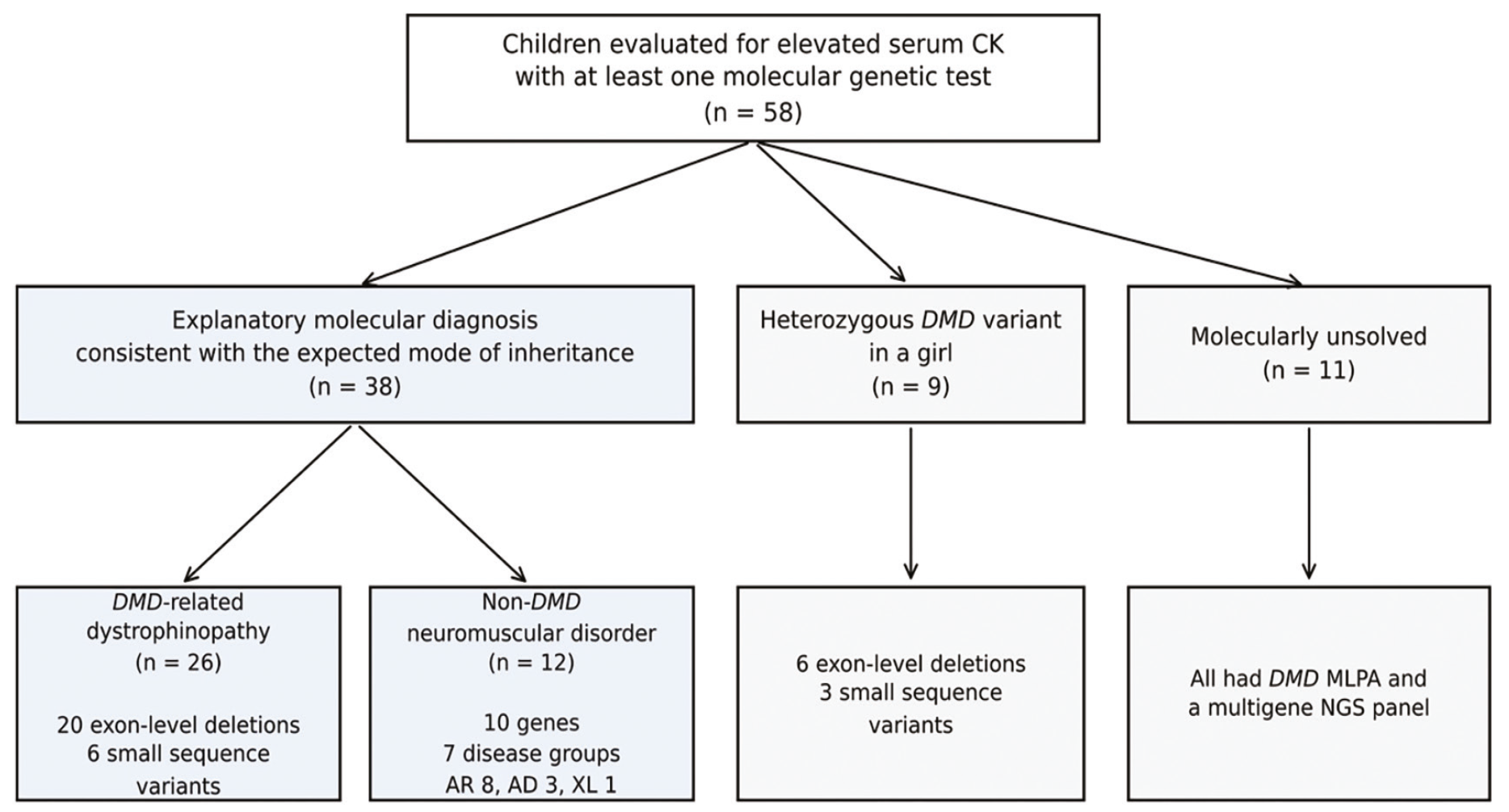
Classification of the cohort according to molecular results.
Table 1. Demographic and laboratory characteristics of the cohort by molecular category
Variable | Explanatory molecular diagnosis (n=38) | Heterozygous DMD variant (n=9) | Molecularly unsolved (n=11) | Whole cohort (n=58) |
|---|---|---|---|---|
Sex, male/female | 34/4 | 0/9 | 8/3 | 42/16 |
Age, years | 2 (1-6) | 9 (5-14) | 1 (0-11) | 4 (1-10) |
Peak CK, U/L | 7,249 (1,468-11,385) | 3,005 (2,071-4,404) | 1,030 (546-3,800) | 4,314 (1,339-9,892) |
Peak AST, U/L | 176 (62-270) | 82 (53-98) | 52 (50-94) | 111 (51-254 |
Peak ALT, U/L | 167 (62-271) | 65 (46-87) | 55 (30-101) | 105 (48-263) |
Continuous variables are presented as median (interquartile range) of peak recorded values. Age is given in completed years, so that children under one year of age contribute a value of 0 to the numerical summaries. “Molecularly unsolved” denotes patients in whom no explanatory molecular finding was identified with the investigations performed. AST and ALT values were available for 54 patients; summaries were calculated using available observations. The institutional upper reference limit for CK was 200 U/L. CK: Creatine kinase, AST: Aspartate aminotransferase, ALT: Alanine aminotransferase.
Distribution of Molecular Diagnoses
Among the 38 patients with an explanatory molecular diagnosis, 26 (68.4%; 95% CI 52.5-80.9) had a DMD-related dystrophinopathy and 12 (31.6%; 95% CI 19.1-47.5) had a non-DMD neuromuscular disorder. Girls with a heterozygous DMD variant were analysed as a separate molecular category and were not included in this denominator. The distribution of diagnoses is shown in .
Table 2. Distribution of explanatory molecular diagnoses (n=38)
Diagnostic group | n (%) | Peak CK, U/L |
|---|---|---|
DMD-related dystrophinopathy | 26 (68.4) | 9,519 (6,667) |
Non-DMD neuromuscular disorder | 12 (31.6) | 1,558 (896-2,815) |
Limb-girdle muscular dystrophy | 6 (15.8) | NC |
Bethlem myopathy | 1 (2.6) | NC |
Emery-Dreifuss muscular dystrophy | 1 (2.6) | NC |
Pompe disease | 1 (2.6) | NC |
McArdle disease | 1 (2.6) | NC |
Myotonia congenita | 1 (2.6) | NC |
Periodic paralysis | 1 (2.6) | NC |
Peak CK values are presented as median (interquartile range) in U/L. NC: Not calculated; CK summaries are not presented for individual non-DMD subgroups because of the small subgroup sizes. CK: Creatine kinase.
DMD Variant Spectrum in Dystrophinopathy
Of the 26 patients with DMD-related dystrophinopathy, 20 (76.9%; 95% CI 57.9-89.0) carried an exon-level deletion and 6 (23.1%) carried a small sequence variant; no exon-level duplication was identified. Of the 20 deletions, 13 (65.0%; 95% CI 43.3-81.9) partially or completely overlapped exons 45-52. Exon 48 was the single exon most frequently included within a deleted interval, followed by exons 46, 47, 49 and 50 (). Of the 19 deletions that could be classified, 11 were predicted to be out-of-frame and 8 in-frame. Peak CK values were lower in patients with an in-frame deletion (median 3,466 U/L, range 611-12,046) than in those with an out-of-frame deletion (median 11,385 U/L, range 5,107-21,221). At the first genetic evaluation, five of the eight patients with an in-frame deletion and four of the nine patients with an out-of-frame deletion and a documented evaluation were asymptomatic; all four of the latter were aged 3 years or younger. Two patients, twin brothers, carried the same splice-site variant. In two patients the deletion was initially detected by chromosomal microarray and subsequently confirmed at the exon level by MLPA. Patient-level variant and clinical data are presented in .
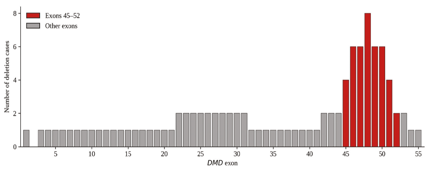
Distribution of DMD exonic deletions in dystrophinopathy cases (n=20). Exons within the 45-52 interval are shown in red, as indicated in the legend. The y-axis indicates the number of deletion cases involving the respective exon; a multi-exon deletion contributes to more than one exon column.
Table 3. Patient-level variant and clinical characteristics of DMD-related dystrophinopathy cases (n=26)
Case | Sex | Age, years | Peak CK, U/L | Exon range/HGVS | Variant type | Overlaps exons 45–52 | Detection method | Reading frame | Clinical findings at first evaluation |
|---|---|---|---|---|---|---|---|---|---|
D1 | M | 1 | 9,581 | Exon 1 deletion | Exonic deletion | No | MLPA | NA | Asymptomatic (calf muscle pseudohypertrophy) |
D2 | M | 2 | 7,122 | Exon 3-7 deletion | Exonic deletion | No | MLPA | Out-of-frame | Gowers sign, calf muscle pseudohypertrophy, myalgia; other: arthralgia |
D3 | M | <1 | 9,457 | Exon 8-44 deletion | Exonic deletion | No | MLPA | Out-of-frame | Asymptomatic; other: facial dysmorphism |
D4 | M | 1 | 21,221 | Exon 22-31 deletion | Exonic deletion | No | MLPA | Out-of-frame | Asymptomatic |
D5 | M | 13 | 611 | Exon 42-44 deletion | Exonic deletion | No | MLPA | In-frame | Asymptomatic; other: intellectual disability, seizure, scoliosis, facial dysmorphism |
D6 | M | 4 | 6,667 | Exon 45-48 deletion | Exonic deletion | Yes | MLPA | In-frame | Asymptomatic |
D7 | M | 5 | 1,468 | Exon 45-51 deletion | Exonic deletion | Yes | MLPA | In-frame | Gowers sign, gait disturbance, toe walking, myalgia |
D8 | M | 12 | 8,321 | Exon 45-47 deletion | Exonic deletion | Yes | MLPA | In-frame | Proximal lower limb muscle weakness, Gowers sign, calf muscle pseudohypertrophy; other: facial dysmorphism |
D9 | M | <1 | 1,396 | Exon 45-47 deletion | Exonic deletion | Yes | MLPA | In-frame | Asymptomatic |
D10 | M | 1 | 11,385 | Exon 46-47 deletion | Exonic deletion | Yes | MLPA | Out-of-frame | Gowers sign, calf muscle pseudohypertrophy; other: arthralgia |
D11 | M | <1 | 17,860 | Exon 46-50 deletion | Exonic deletion | Yes | MLPA | Out-of-frame | Not documented |
D12 | M | 18 | 8,246 | Exon 48-50 deletion | Exonic deletion | Yes | MLPA | Out-of-frame | Loss of ambulation; other: treated left ventricular systolic dysfunction |
D13 | M | 3 | 20,909 | Exon 48-50 deletion | Exonic deletion | Yes | MLPA | Out-of-frame | Asymptomatic |
D14 | M | 2 | 5,464 | Exon 48 deletion | Exonic deletion | Yes | MLPA | In-frame | Gowers sign |
D15 | M | 3 | 19,069 | Exon 48-52 deletion | Exonic deletion | Yes | MLPA | Out-of-frame | Not documented |
D16 | M | 2 | 12,046 | Exon 48-51 deletion | Exonic deletion | Yes | MLPA | In-frame | Asymptomatic |
D17 | M | 6 | 15,720 | Exon 51 deletion | Exonic deletion | Yes | MLPA | Out-of-frame | Gowers sign, calf muscle pseudohypertrophy |
D18 | M | 2 | 1,379 | Exon 52-53 deletion | Exonic deletion | Yes | MLPA | In-frame | Asymptomatic; other: facial dysmorphism |
D19 | M | <1 | 9,892 | Exon 53-54 deletion | Exonic deletion | No | MLPA | Out-of-frame | Asymptomatic |
D20 | M | 2 | 5,107 | Exon 55 deletion | Exonic deletion | No | MLPA | Out-of-frame | Toe walking, calf muscle pseudohypertrophy |
D21 | M | 1 | 13,300 | c.10223+1G>A | Small sequence variant | NA | Sequence analysis | NA | Delayed ability to walk |
D22 | M | <1 | 13,647 | c.7309+5G>C | Small sequence variant | NA | Sequence analysis | NA | Asymptomatic |
D23 | M | 4 | 10,138 | c.5899C>T (p.Arg1967Ter) | Small sequence variant | NA | Sequence analysis | NA | Gowers sign, toe walking, calf muscle pseudohypertrophy, exercise intolerance |
D24 | M | 7 | 7,686 | c.5287C>T (p.Arg1763Ter) | Small sequence variant | NA | Sequence analysis | NA | Difficulty climbing stairs, delayed ability to walk, exercise intolerance; other: delayed speech and language development |
D25 | M | 1 | 9,416 | c.2786_2788delinsG | Small sequence variant | NA | Sequence analysis | NA | Asymptomatic |
D26 | M | 1 | 14,760 | c.10223+1G>A | Small sequence variant | NA | Sequence analysis | NA | Delayed ability to walk |
HGVS descriptions are presented as recorded in the available clinical laboratory reports; protein consequences are given in parentheses where reported. Small sequence variants comprise nonsense, frameshift, indel and splice-site changes. Ages below one year are shown as <1. M, male; MLPA, multiplex ligation-dependent probe amplification; NA, not applicable (overlap with exons 45-52 was assessed only for exon-level deletions). In cases D5 and D18 the deletion was initially detected by chromosomal microarray and confirmed by MLPA. Cases D21 and D26, who carried the same splice-site variant, are twin brothers. Reference transcripts, ACMG/AMP evidence codes and segregation analyses were not available in the source records. The reading frame was predicted for exon-level deletions from the coding lengths of the deleted exons; the exon 1 deletion was not classified, and NA in this column denotes small sequence variants. Clinical findings recorded at the first genetic evaluation are given as Human Phenotype Ontology terms in a fixed order (weakness, Gowers sign, motor and gait findings, muscle bulk, myalgia and exercise intolerance, myotonia, contractures), followed, after “other”, by findings in other organ systems (developmental and growth, neurological, cardiac dysfunction, spinal and chest deformities, arthralgia and facial dysmorphism). Patients without any neuromuscular complaint or finding are designated asymptomatic, irrespective of findings in other organ systems; isolated calf muscle pseudohypertrophy, given in parentheses, did not classify a patient as symptomatic. Findings recorded as equivocal were considered present (Gowers sign in cases D10 and D14 and calf muscle pseudohypertrophy in case D20). Not documented, no record of the first evaluation available. CK: Creatine kinase; HGVS: Human Genome Variation Society.
Non-DMD Molecular Diagnoses
The 12 non-DMD diagnoses were identified in 10 different genes, and only CAPN3 was represented more than once. They comprised seven distinct disease groups: LGMD (n=6), Bethlem myopathy (n=1), Emery-Dreifuss muscular dystrophy (n=1), Pompe disease (n=1), McArdle disease (n=1), myotonia congenita (n=1) and periodic paralysis (n=1), and three modes of inheritance, with eight autosomal recessive diagnoses (GAA, CAPN3 in three patients, SGCA, TCAP, POMT1, PYGM), three autosomal dominant diagnoses (COL6A1, CLCN1, SCN4A) and one X-linked diagnosis (EMD). This distribution illustrates the potential genetic and mechanistic heterogeneity of non-DMD causes in this setting, although the small number of patients precludes firm conclusions regarding their relative frequencies. Nine of the 12 patients had a neuromuscular complaint or finding at the first genetic evaluation. Variant-level and clinical data are presented in .
Table 4. Variant-level and clinical characteristics of non-DMD molecular diagnoses (n=12)
Case | Sex | Gene | Variant (HGVS) | Zygosity | Inh. | Disease | Peak CK, U/L | Detection method | Clinical findings at first evaluation |
|---|---|---|---|---|---|---|---|---|---|
P1 | M | GAA | c.1655T>C (p.Leu552Pro); c.2741delinsCAG (p.Gln914ProfsTer30) | Two variants; phase not determined | AR | Pompe disease | 942 | NGS panel | Asymptomatic |
P2 | F | CAPN3 | c.1381C>T (p.Arg461Cys) | Homozygous | AR | LGMD R1 calpain3-related | 1,777 | NGS panel | Myalgia, exercise intolerance |
P3 | M | COL6A1 | c.824G>A (p.Gly275Glu) | Heterozygous | AD | Bethlem myopathy | 423 | NGS panel | Proximal lower limb muscle weakness, frequent falls, tight Achilles tendon |
P4 | M | POMT1 | c.598G>C (p.Ala200Pro) | Homozygous | AR | LGMD R11 POMT1-related | 7,376 | NGS panel | Delayed ability to walk; other: delayed speech and language development, growth delay, facial dysmorphism |
P5 | F | SGCA | c.739G>A (p.Val247Met) | Homozygous | AR | LGMD R3 α-sarcoglycan-related | 4,224 | NGS panel | Asymptomatic |
P6 | M | CLCN1 | c.501C>G (p.Phe167Leu) | Heterozygous | AD | Myotonia congenita | 268 | NGS panel | Myotonia |
P7 | M | EMD | c.506_507del (p.Pro169ArgfsTer60) | Hemizygous | XL | Emery-Dreifuss muscular dystrophy | 851 | NGS panel | Difficulty climbing stairs, frequent falls, toe walking, exercise intolerance |
P8 | M | TCAP | c.90_91del (p.Ser31HisfsTer11) | Homozygous | AR | LGMD R7 telethonin-related | 1,339 | Exome sequencing | Limb-girdle muscle weakness, Gowers sign, limited elbow movement, limitation of knee mobility; other: kyphosis, pectus carinatum |
P9 | M | SCN4A | c.2111C>T (p.Thr704Met) | Heterozygous | AD | Periodic paralysis | 1,163 | Sequence analysis | Limb-girdle muscle weakness, episodic flaccid weakness, skeletal muscle atrophy |
P10 | F | CAPN3 | c.1469G>A (p.Arg490Gln) | Homozygous | AR | LGMD R1 calpain3-related | 1,786 | NGS panel | Toe walking; other: facial dysmorphism |
P11 | M | PYGM | c.2262del | Homozygous | AR | McArdle disease | 1,879 | Sequence analysis | Asymptomatic |
P12 | F | CAPN3 | c.245C>T (p.Pro82Leu); c.1621C>T (p.Arg541Trp) | Compound heterozygous | AR | LGMD R1 calpain3-related | 3,751 | NGS panel | Myalgia |
HGVS descriptions are presented as recorded in the available clinical laboratory reports; gene symbols are italicised and protein consequences are given in parentheses where reported. M: Male, F: Female, AR: Autosomal recessive, AD: Autosomal dominant, XL: X-linked, Inh.: Inheritance, LGMD: Limb-girdle muscular dystrophy, NGS: Next-generation sequencing, CK: Creatine kinase, HGVS: Human Genome Variation Society. Reference transcripts, ACMG/AMP evidence codes and segregation analyses were not available in the source records. Clinical findings are reported as in . LGMD subtypes are named according to the current nomenclature (). In case P1, two GAA variants were reported and the phase was not determined; echocardiography showed no cardiomyopathy. | |||||||||
Heterozygous DMD Variants in Girls
Nine girls carried a heterozygous DMD variant, comprising six exon-level deletions and three small sequence variants. Standardised neuromuscular examination data, including muscle strength assessment, were not available for these patients; they were therefore analysed as a separate molecular category, and no inference regarding their clinical status was made. Four girls had a neuromuscular complaint or finding at the first genetic evaluation. Peak recorded CK values in this group ranged from 271 to 12,768 U/L ().
Table 5. Molecular, laboratory and clinical characteristics of girls with a heterozygous DMD variant (n=9)
Case | Variant (HGVS) | Zygosity | Peak CK, U/L | Detection method | Clinical findings at first evaluation |
|---|---|---|---|---|---|
T1 | c.2104G>T (p.Glu702Ter) | Heterozygous | 2,071 | NGS panel | Exercise intolerance |
T2 | Exon 50 deletion | Heterozygous | 2,614 | MLPA | Asymptomatic; other: arthralgia |
T3 | Exon 48-50 deletion | Heterozygous | 3,005 | MLPA | Asymptomatic |
T4 | Exon 10-11 deletion | Heterozygous | 1,170 | MLPA | Proximal lower limb muscle weakness, Gowers sign |
T5 | c.5612dup (p.Ala1872GlyfsTer16) | Heterozygous | 3,199 | NGS panel | Limb-girdle muscle weakness |
T6 | c.519_522del (p.His174ValfsTer33) | Heterozygous | 7,795 | NGS panel | Asymptomatic |
T7 | Exon 42-44 deletion | Heterozygous | 271 | MLPA | Asymptomatic |
T8 | Exon 10-43 deletion | Heterozygous | 4,404 | MLPA | Gait disturbance, exercise intolerance; other: global developmental delay, spasticity, ophthalmoplegia, tremor, dysphagia, scoliosis |
T9 | Exon 45-47 deletion | Heterozygous | 12,768 | MLPA | Asymptomatic |
HGVS descriptions are presented as recorded in the available clinical laboratory reports; protein consequences are given in parentheses where reported. MLPA: Multiplex ligation-dependent probe amplification; NGS: Next-generation sequencing, CK: Creatine kinase, HGVS: Human Genome Variation Society. Reference transcripts, ACMG/AMP evidence codes, segregation analyses and standardised neuromuscular examination data were not available in the source records. Clinical findings are reported as in . | |||||
Peak Serum CK Values
The median peak CK in the whole cohort was 4,314 U/L (IQR 1,339-9,892; range 268-45,550). Median values were 7,249 U/L (,385) in patients with an explanatory molecular diagnosis, 3,005 U/L (,404) in girls with a heterozygous DMD variant and 1,030 U/L (800) in molecularly unsolved patients. Within the group with an explanatory molecular diagnosis, median peak CK was 9,519 U/L (,647) in dystrophinopathy and 1,558 U/L (815) in non-DMD disorders, and within dystrophinopathy it was 8,889 U/L (,883) in patients with an exonic deletion and 11,719 U/L (,647) in those with a small sequence variant. Peak recorded CK values showed substantial descriptive overlap among molecular categories (). No inferential statistical comparison between categories was performed.
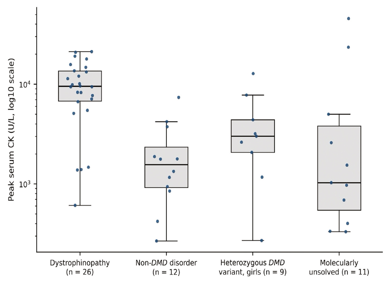
Distribution of peak serum creatine kinase across molecular categories (log10 scale). Boxes indicate the interquartile range, the line within the box the median, and whiskers the most extreme values within 1.5×IQR; points represent individual patients. No inferential comparison was performed between categories.
Clinical Features and Course of CK Elevation
Clinical features are summarised in . The context in which CK elevation was first detected was documented in 46 patients: an incidental measurement in 17, investigation of elevated transaminases in 16, evaluation of muscle symptoms in 8 and other circumstances in 5. A neuromuscular complaint or finding was recorded at the first genetic evaluation in 35 of 56 patients. At least two CK measurements were available in 43 patients; CK elevation was persistent in 29 and transient in 2, and could not be assessed in 27 because fewer than two measurements, or only measurements less than 8 weeks apart, were available. Both patients with transient elevation were molecularly unsolved, whereas all non-DMD diagnoses with repeated measurements showed persistent elevation.
Table 6. Clinical features at first evaluation and course of CK elevation by molecular category
Variable | Dystrophinopathy (n=26) | Non-DMD disorder (n=12) | Heterozygous DMD variant (n=9) | Molecularly unsolved (n=11) | Whole cohort (n=58) |
|---|---|---|---|---|---|
Context of detection documented | 18 | 11 | 7 | 10 | 46 |
Incidental measurement | 7 | 3 | 4 | 3 | 17 |
Investigation of elevated transaminases | 8 | 3 | 2 | 3 | 16 |
Evaluation of muscle symptoms | 2 | 4 | 0 | 2 | 8 |
Other | 1 | 1 | 1 | 2 | 5 |
Examination at first evaluation documented | 24 | 12 | 9 | 11 | 56 |
Neuromuscular complaint or finding present | 12 | 9 | 4 | 10 | 35 |
≥2 CK measurements available | 16 | 10 | 8 | 9 | 43 |
CK course: persistent | 11 | 10 | 5 | 3 | 29 |
CK course: transient | 0 | 0 | 0 | 2 | 2 |
CK course: not assessable | 15 | 2 | 4 | 6 | 27 |
Consanguinity documented | 20 | 11 | 6 | 8 | 45 |
Consanguinity present (first cousins) | 5 (1) | 5 (4) | 1 (0) | 4 (3) | 15 (8) |
Family history documented | 20 | 11 | 8 | 8 | 47 |
Family history of neuromuscular disease | 8 | 5 | 3 | 2 | 18 |
Values are numbers of patients. Variables were extracted retrospectively from clinical records; “documented” denotes the number of patients in whom the variable was recorded. A neuromuscular complaint or finding denotes weakness, Gowers sign, gait disturbance, myalgia, exercise intolerance, myotonia, hypotonia or motor delay recorded at the first genetic evaluation; equivocal findings were considered present. CK course: persistent, CK above the upper reference limit on at least two occasions more than 8 weeks apart without subsequent normalisation; transient, normalisation during follow-up; not assessable, fewer than two measurements or measurements less than 8 weeks apart. The number of first-cousin unions is given in parentheses. CK: Creatine kinase.
Discussion
In this single-centre cohort of children evaluated for elevated serum CK, DMD-related dystrophinopathy was the predominant molecular diagnosis, but it did not account for the whole of the explained aetiology. Approximately one third of molecular diagnoses were attributable to disorders unrelated to DMD, distributed across several genes, inheritance patterns and disease mechanisms rather than concentrated in a small number of alternative loci. Taken together, these observations indicate that the molecular basis of unexplained CK elevation in childhood is broader than dystrophinopathy alone, and that the differential diagnosis should be framed accordingly when a child is referred for genetic evaluation.
These findings do not imply that every child with elevated CK requires genetic testing. In this cohort, CK elevation was most often detected incidentally or during investigation of elevated transaminases, and persistence could not be assessed in almost half of the patients; conversely, more than half had a neuromuscular complaint or finding at the first genetic evaluation, all non-DMD diagnoses with repeated measurements showed persistent elevation, consanguinity was frequent among the non-DMD diagnoses, and the only two patients whose CK normalised remained molecularly unsolved. These observations are consistent with the stepwise, phenotype-driven approach proposed in paediatric series (,), in which the elevation is first confirmed on repeated measurement, with recent exercise, trauma or intercurrent illness excluded (), and history, family history, physical examination and targeted laboratory tests then determine whether, and which, genetic test is indicated. In the two channelopathies in this cohort, myotonia and episodic weakness, rather than the CK level, pointed to the diagnosis.
The relevance of the non-DMD diagnoses lies less in their number than in their distribution. The 12 diagnoses involved 10 different genes, spanning seven disease groups and three modes of inheritance, and only CAPN3 was represented more than once. This pattern illustrates the breadth of the differential diagnosis of paediatric CK elevation and is consistent with the marked genetic heterogeneity reported in NGS-based series of inherited myopathies and hyperCKemia (,,). Because the number of patients is limited, these findings should not be taken to establish the relative frequencies of individual disorders; they suggest, rather, that a diagnostic approach directed only at the most common cause may leave a clinically meaningful minority unexplained. The predominance of autosomal recessive LGMD subtypes among our non-DMD diagnoses is consistent with a previous Turkish series in which all molecularly confirmed LGMD subtypes were autosomal recessive and consanguinity was frequent ().
Four diagnoses fell outside the category of muscular dystrophy altogether: two metabolic myopathies (Pompe disease and McArdle disease) and two muscle channelopathies (myotonia congenita and periodic paralysis). Their identification is clinically relevant because disorders of this kind require different monitoring and, in the case of Pompe disease, disease-specific enzyme replacement therapy, for which a positive effect at group level, with considerable inter-individual variability in response, has been reported in children with non-classic disease during long-term follow-up (). Recognising such conditions promptly is therefore of practical importance in a child whose only initial abnormality is a raised CK, and metabolic disorders have similarly been reported in paediatric series of CK elevation (,).
Although peak CK values were generally higher in dystrophinopathy than in other categories, the distributions overlapped substantially, and several non-DMD diagnoses were identified at comparatively modest CK concentrations. No inferential comparison was undertaken, so no discriminatory value can be attributed to CK in this cohort. From a practical standpoint, the magnitude of CK elevation should not by itself be used to prioritise or to exclude particular genetic diagnoses, and it is best interpreted alongside the clinical picture and family history, particularly as paediatric CK reference limits vary with age and sex ().
Our dystrophinopathy findings are consistent with previous work. Exonic deletions predominated and most overlapped the central exon 45-52 hotspot, in line with the predominance and central clustering of deletions described in large dystrophinopathy cohorts (,). The absence of exon-level duplications contrasts with those cohorts, in which exon-level duplications accounted for 13% of registered patients and 10.7% of molecularly diagnosed patients, respectively (,), and may be related to the limited number of cases. Peak CK values were lower in patients with an in-frame deletion than in those with an out-of-frame deletion. Symptom status at the first evaluation, however, was not clearly different between the two small groups and largely reflected age, because most patients were evaluated in early childhood, whereas the Duchenne and Becker phenotypes are defined by the subsequent course of ambulation (). These genotype–phenotype relationships are therefore descriptive and require longitudinal follow-up. The predominance of dystrophinopathy among our molecular diagnoses accords with paediatric series of CK elevation, in which dystrophinopathies were the most frequent inherited neuromuscular diagnosis (,,), whereas the joint identification of dystrophinopathy and non-DMD diagnoses in our cohort may partly reflect the fact that it was not restricted by symptom status, in contrast to series confined to asymptomatic or minimally symptomatic individuals, in which overtly symptomatic dystrophinopathy is underrepresented or excluded by design (,,).
Girls with a heterozygous DMD variant were analysed as a separate molecular category. Standardised neuromuscular assessment, including muscle strength testing, was not available for these patients. In a survey of 32 confirmed female carriers of DMD variants reported alongside a population-based CK screening programme, only half had elevated CK, and CK values varied even among carriers of the same variant (); the clinical status of a carrier cannot be inferred from the molecular finding or the CK level alone. Assigning these girls to either the diagnosed or the unexplained group would therefore have misrepresented their status and altered the denominator on which the aetiological proportions were calculated.
The main strengths of this study are the joint description of DMD-related and non-DMD diagnoses within a single referred cohort, its molecular classification and its transparency. All explanatory diagnoses were based on molecularly confirmed pathogenic or likely pathogenic variants. Results were assigned to three mutually exclusive categories with explicit denominators; heterozygous DMD variants in girls were analysed separately rather than merged with disease diagnoses; findings are reported at the level of individual patients and variants rather than as aggregate counts; and molecularly unsolved patients were interpreted conservatively rather than as true negatives.
Study Limitations
Several limitations should be considered. The study is retrospective and single-centre, and referral patterns to a tertiary genetics clinic are likely to have influenced the composition of the cohort. Genetic testing was performed according to clinical indication rather than a uniform protocol, so the extent of investigation differed between patients; although all molecularly unsolved patients had undergone both MLPA and a multigene NGS panel, none had undergone whole-exome or genome sequencing, and deep intronic variants, repeat expansions and genes not covered by the panels may therefore have been missed. Pathogenicity classifications were taken from the final clinical laboratory reports and cross-checked against ClinVar, but an independent ACMG/AMP re-scoring was not undertaken, and one variant accepted as explanatory (CLCN1 p.Phe167Leu) has conflicting classifications in ClinVar. Clinical features were extracted retrospectively from routine records, standardised muscle strength assessment was not available, and persistence of CK elevation could not be assessed in almost half of the patients, which limits genotype–phenotype correlation. Because eligibility was based on a documented peak value rather than on demonstrated persistence, transient causes of CK elevation, such as recent exercise, trauma or viral myositis, cannot be excluded, particularly among molecularly unsolved patients. Consanguinity was not documented in 13 patients. The cohort size also limits the precision of the proportions reported.
Conclusion
DMD-related dystrophinopathies represented the majority of explanatory molecular diagnoses in this selected paediatric referral cohort. However, non-DMD diagnoses encompassed multiple genes, inheritance patterns and disease mechanisms, including clinically actionable metabolic and channel disorders. These findings illustrate that, once persistent elevation has been confirmed and the history, family history and examination have been evaluated, the evaluation of unexplained paediatric CK elevation should extend beyond DMD when initial dystrophinopathy testing is unrevealing or the phenotype is atypical. Prospective multicentre studies using uniform testing and standardised phenotyping are required to better define the diagnostic spectrum and genotype–phenotype relationships.
Ethics
Ethics Committee Approval: The study was approved by the Non-Interventional Clinical Research Ethics Committee of Samsun University (protocol code: GOKAEK 2026/8/12, date of approval: 6 May 2026).
Informed Consent: The requirement for informed consent was waived by the ethics committee because of the retrospective design and the use of anonymised data.
Footnotes
Authorship Contributions
Concept: E.S., Design: E.S., A.S., Data Collection or Processing: E.S., Ü.A., Analysis or Interpretation: E.S., A.U., Literature Search: E.S., Critical Review: E.S., G.A., A.S., N.A., Ü.A., A.U., Writing: E.S.
Conflict of Interest
No conflict of interest was declared by the authors.
Financial Disclosure
The authors declared that this study received no financial support.
References
Fechner A, Willenberg A, Ziegelasch N, Merkenschlager A, Kiess W, Vogel M. Creatine kinase serum levels in children revisited: new reference intervals from a large cohort of healthy children and adolescents. Clin Chim Acta. 2024;560:119726.
Prelle A, Tancredi L, Sciacco M, Chiveri L, Comi GP, Battistel A, et al. Retrospective study of a large population of patients with asymptomatic or minimally symptomatic raised serum creatine kinase levels. J Neurol. 2002;249:305-11.
Gemelli C, Traverso M, Trevisan L, Fabbri S, Scarsi E, Carlini B, et al. An integrated approach to the evaluation of patients with asymptomatic or minimally symptomatic hyperCKemia. Muscle Nerve. 2022;65:96-104.
Han S, Xu H, Zheng J, Sun J, Feng X, Wang Y, et al. Population-wide Duchenne muscular dystrophy carrier detection by CK and molecular testing. Biomed Res Int. 2020;2020:8396429.
Özkan Kart P, Acar Arslan E. A retrospective evaluation of pediatric patients with symptomatic hyperCKemia. Turk Arch Pediatr. 2026;61:230-40.
Al-Ghamdi F, Darras BT, Ghosh PS. Spectrum of neuromuscular disorders with hyperCKemia from a tertiary care pediatric neuromuscular center. J Child Neurol. 2018;33:389-96.
Monteiro C, Garrido C, Pereira Â, Dias A, Costa M, Magalhães C, et al. Multicentric study on the diagnostic of neuromuscular diseases in children with high creatinine phosphokinase levels. Children (Basel). 2024;11:1462.
Okubo M, Goto K, Komaki H, Nakamura H, Mori-Yoshimura M, Hayashi YK, et al. Comprehensive analysis for genetic diagnosis of dystrophinopathies in Japan. Orphanet J Rare Dis. 2017;12:149.
Zhao L, Shi Y, Hu C, Zhou S, Li H, Zhang L, et al. Comprehensive analysis of 2097 patients with dystrophinopathy based on a database from 2011 to 2021. Orphanet J Rare Dis. 2024;19:311.
Carter JC, Sheehan DW, Prochoroff A, Birnkrant DJ. Muscular dystrophies. Clin Chest Med. 2018;39:377-89.
Bouchard C, Tremblay JP. Limb-girdle muscular dystrophies classification and therapies. J Clin Med. 2023;12:4769.
Falsaperla R, Praticò AD, Ruggieri M, Parano E, Rizzo R, Corsello G, et al. Congenital muscular dystrophy: from muscle to brain. Ital J Pediatr. 2016;42:78.
Rubegni A, Malandrini A, Dosi C, Astrea G, Baldacci J, Battisti C, et al. Next-generation sequencing approach to hyperCKemia: a 2-year cohort study. Neurol Genet. 2019;5:e352.
Marti P, Pitarch-Castellano I, Muelas N, Azorín I, Fores L, Vilchez R, et al. Asymptomatic hyperCKemia in the pediatric population: a prospective study utilizing next-generation sequencing and ancillary tests. Neurology. 2025;104:e210116.
Invernizzi F, Izzo R, Colangelo I, Legati A, Zanetti N, Garavaglia B, et al. NGS-based genetic analysis in a cohort of Italian patients with suspected inherited myopathies and/or hyperCKemia. Genes (Basel). 2023;14:1393.
Aires Martins I, Baptista de Lima J, Coelho MP, Bandeira A, Martins E, Correia J. Pediatric hyperCKemia: a 13-year retrospective study and predictors of neuromuscular disease and metabolic myopathy. Eur J Pediatr. 2026;185:250.
Yoldaş Çelik M, Köşeci B. Identifying inherited metabolic disorders in children: the role of elevated creatine kinase. BMC Pediatr. 2025;25:612.
von Elm E, Altman DG, Egger M, Pocock SJ, Gøtzsche PC, Vandenbroucke JP; STROBE Initiative. The Strengthening the Reporting of Observational Studies in Epidemiology (STROBE) statement: guidelines for reporting observational studies. J Clin Epidemiol. 2008;61:344-9.
Richards S, Aziz N, Bale S, Bick D, Das S, Gastier-Foster J, et al. Standards and guidelines for the interpretation of sequence variants: a joint consensus recommendation of the American College of Medical Genetics and Genomics and the Association for Molecular Pathology. Genet Med. 2015;17:405-24.
Sarıkaya Uzan G, Yılmaz Uzman C, Çinleti T, Günay Ç, Ülgenalp A, Hız Kurul S, et al. Molecular diagnosis of limb-girdle muscular dystrophy using next-generation sequencing panels. Mol Syndromol. 2024;15:14-21.
van der Meijden JC, Kruijshaar ME, Harlaar L, Rizopoulos D, van der Beek NAME, van der Ploeg AT. Long-term follow-up of 17 patients with childhood Pompe disease treated with enzyme replacement therapy. J Inherit Metab Dis. 2018;41:1205-14.